Structure and Function of Eye

When surveyed about the five senses — sight, hearing, taste, smell and touch — people consistently report that their eyesight is the mode of perception they value (and fear losing) most.
Despite this, many people don't have a good understanding of the anatomy of the eye, how vision works, and health problems that can affect the eye.

Read on for a basic description and explanation of the structure (anatomy) of your eyes and how they work (function) to help you see clearly and interact with your world.
Structure of eye:
Anterior chamber: 
                         The region of the eye between the cornea and the lens that contains aqueous humor.
Aqueous humor: 
                        The fluid produced in the eye.
Bruch's membrane: 
                        Located in the retina between the choroid and the retinal pigmented epithelium (RPE) layer; provides support to the retina and functions as the 'basement' membrane of the RPE layer.

Ciliary body: 
            Part of the eye, above the lens, that produces the aqueous humor.
Choroid: 
           Layer of the eye behind the retina, contains blood vessels that nourish the retina.
Cones: 
           The photoreceptor nerve cells present in the macula and concentrated in the fovea (the very center of the macula); enable people to see fine detail and color.
Cornea:
           The outer, transparent structure at the front of the eye that covers the iris, pupil and anterior chamber; it is the eye's primary light-focusing structure.
Drusen: 
            Deposits of yellowish extra cellular waste products that accumulate within and beneath the retinal pigmented epithelium (RPE) layer.
Fovea: 
            The pit or depression at the center of the macula that provides the greatest visual acuity.
Iris:  
         The colored ring of tissue behind the cornea that regulates the amount of light entering the eye by adjusting the size of the pupil.
Lens: 
           The transparent structure suspended behind the iris that helps to focus light on the retina; it primarly provides a fine-tuning adjustment to the primary focusing structure of the eye, which is the cornea.
Macula: 
            The portion of the eye at the center of the retina that processes sharp, clear straight-ahead vision.
Optic nerve: 
            The bundle of nerve fibers at the back of the eye that carry visual messages from the retina to the brain.
Photoreceptors: 
           The light sensing nerve cells (rods and cones) located in the retina.
Pupil: 
           The adjustable opening at the center of the iris through which light enters the eye.
Retina: 
             The light sensitive layer of tissue that lines the back of the eye.
Retinal Pigmented Epithelium (RPE):
              A layer of cells that protects and nourishes the retina, removes waste products, prevents new blood vessel growth into the retinal layer and absorbs light not absorbed by the photoreceptor cells; these actions prevent the scattering of the light and enhance clarity of vision.
Rods: 
            Photoreceptor nerve cells in the eyes that are sensitive to low light levels and are present in the retina, but outside the macula.
Sclera: 
             The tough outer coat that protects the entire eyeball.
Trabecular meshwork: 
             Spongy tissue located near the cornea through which aqueous humor flows out of the eye.
Vitreous:
             Clear jelly-like substance that fills the eye from the lens to the back of the eye.
     
  How The Eye Works

  1. Light is focused primarily by the cornea — the clear front surface of the eye, which acts like a camera lens.
  2. The iris of the eye functions like the diaphragm of a camera, controlling the amount of light reaching the back of the eye by automatically adjusting the size of the pupil (aperture).
  3. The eye's crystalline lens is located directly behind the pupil and further focuses light. Through a process called accommodation, this lens helps the eye automatically focus on near and approaching objects, like an autofocus camera lens.
  4. Light focused by the cornea and crystalline lens (and limited by the iris and pupil) then reaches the retina — the light-sensitive inner lining of the back of the eye. The retina acts like an electronic image sensor of a digital camera, converting optical images into electronic signals. The optic nerve then transmits these signals to the visual cortex — the part of the brain that controls our sense of sight.
  5. In a number of ways, the human eye works much like a digital camera:


Function of nephrone

Nephron
It is the functional unit of kidneys. The nephron is the basic structural and functional unit of the kidney. Its chief function is to regulate the concentration of water and soluble substances like sodium salts by filtering the blood, reabsorbing what is needed and excreting the rest as urine. A nephron eliminates wastes from the body, regulates blood volume and blood pressure, controls levels of electrolytes and metabolites, and regulates blood pH


A nephron is the basic unit of structure in the kidney. A nephron is used separate to water, ions and small molecules from the blood, filter out wastes and toxins, and return needed molecules to the blood. The nephron functions through ultrafiltration. Ultrafiltration occurs when blood pressure forces water and other small molecules through tiny gaps in capillary walls. This substance, lacking the blood cells and large molecules in the bloodstream, is known as an ultrafiltrate. The ultrafiltrate travels through the various loops of the nephron, where water and important molecules are removed, and into a collecting duct which drains into the bladder.

Structure of Nephron

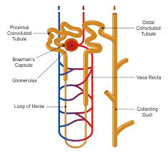
The picture below is of a general nephron. This nephron contains a loop of Henle, so it is a mammalian nephron. While the loop of the nephron is special to mammals, the rest of the structure is seen in all vertebrate animals. The glomerulus is the net of capillaries inside of the glomerular capsule (aka Bowman’s capsule). While the picture shows the glomerular capsule and the rest of the renal tubule look to be the same in the graphic below, they are in fact composed of a wide variety of cell types, intended to extract and retain certain chemicals within the tubules.
Each nephron consists of one main interlobular artery feeding a single renal tubule. Each kidney in a vertebrate has hundreds to millions of nephrons, each of which produces urine and sends it to the bladder. The cells in each nephron are arranged so that the most concentrated cells are at the bottom of the nephron, while the cells at the top are less concentrated. The cells near the exit of the nephron are the most concentrated, and therefore extract as much water as possible from the ultrafiltrate before it is sent to the bladder.
Function of a Nephron
A nephron is responsible for removing waste products, stray ions, and excess water from the blood. The blood travels through the glomerulus, which is surrounded by the glomerular capsule. As the heart pumps the blood, the pressure created pushes small molecules through the capillaries and into the glomerular capsule. This is the, more physical function of the nephron. Next, the ultrafiltrate must travel through a winding series of tubules. The cells in each part of the tube have different molecules that they like to absorb. Molecules to be excreted remain in the tubule, while water, glucose and other beneficial molecules work their way back into the bloodstream. As the ultrafiltrate travels down the tubules, the cells become more and more hypertonic compared to the ultrafiltrate. This causes a maximum amount of water to be extracted from the ultrafiltrate before it exits the nephron. The blood surrounding the nephron returns to the body via the interlobular vein, free of toxins and excess substances. The ultrafiltrate is now urine, and moves via the collecting duct to the bladder, where it will be stored.

Homeostasis in human

Homeostasis
Homeostasis refers to the ability of an organism or environment to maintain stability in spite of changes. The human body is full of examples of homeostasis.


Homeostasis refers to stability, balance, or equilibrium within a cell or the body. It is an organism’s ability to keep a constant internal environment. Homeostasis is an important characteristic of living things. Keeping a stable internal environment requires constant adjustments as conditions change inside and outside the cell. The adjusting of systems within a cell is called homeostatic regulation. Because the internal and external environments of a cell are constantly changing, adjustments must be made continuously to stay at or near the set point (the normal level or range). Homeostasis can be thought of as a dynamic equilibrium rather than a constant, unchanging state.

Common Homeostasis Examples


  1. Humans’ internal body temperature is a great example of homeostasis. When an individual is healthy, his or her body temperature retains a temperature 98.6 degrees Fahrenheit. The body can control temperature by making or releasing heat. 
  2. Glucose is a type of sugar that is found in the bloodstream, but the body must maintain proper glucose levels to ensure that a person remains healthy. When glucose levels get too high, the pancreas releases a hormone known as insulin. If blood glucose levels happen to drop too low, the liver converts glycogen in the blood to glucose again, raising the levels.
  3. When bacteria or viruses that can make you ill get into your body, your lymphatic system kicks in to help maintain homeostasis. It works to fight the infection before it has the opportunity to make you sick, ensuring that you remain healthy.
  4. The maintenance of healthy blood pressure is an example of homeostasis. The heart can sense changes in the blood pressure, causing it to send signals to the brain, which then sends back signals telling the heart how to respond. If blood pressure is too high, naturally the heart should slow down; while if it is too low, the heart wants to speed up.
  5. A human’s body contains chemicals known as acids and bases, and a proper balance of these is required for the body to function optimally. Lungs and kidneys are two of the organ systems that regulate acids and bases within the body. 
  6. More than half of a human’s body weight percentage is water, and maintaining the correct balance of water is an example of homeostasis. Cells that have too much water in them bloat and can even blow up. Cells with too little water can end up shrinking. Your body maintains a proper water balance so that neither of these situations occurs.
  7. Calcium levels in the blood must be maintained at proper levels. The body regulates those levels in an example of homeostasis. When levels decrease, the parathyroid releases hormones. If calcium levels become too high, the thyroid helps out by fixing calcium in the bones and lowering blood calcium levels.
  8. Exercising causes the body to maintain homeostasis by sending lactate to the muscles to give them energy. Over time, this also signals to the brain that it is time to stop exercising, so that the muscles can get the oxygen they need.
  9. The nervous system helps keep homeostasis in breathing patterns. Because breathing is involuntary, the nervous system ensures that the body is getting much needed oxygen through breathing the appropriate amount of oxygen.
  10. When toxins get into your blood, they disrupt your body’s homeostasis. The human body, however, responds by getting rid of these toxins by use of the urinary system. An individual simply urinates the toxins and other nasty things from the blood, restoring homeostasis to the human body.  

Feedback Regulation Loops
The endocrine system plays an important role in homeostasis because hormones regulate the activity of body cells. The release of hormones into the blood is controlled by a stimulus. For example, the stimulus either causes an increase or a decrease in the amount of hormone secreted. Then, the response to a stimulus changes the internal conditions and may itself become a new stimulus. This self-adjusting mechanism is called feedback regulation.
Feedback regulation occurs when the response to a stimulus has an effect of some kind on the original stimulus. The type of response determines what the feedback is called. Negative feedback occurs when the response to a stimulus reduces the original stimulus. Positive feedback occurs when the response to a stimulus increases the original stimulus.
Thermoregulation: A Negative Feedback Loop
Negative feedback is the most common feedback loop in biological systems. The system acts to reverse the direction of change. Since this tends to keep things constant, it allows the maintenance of homeostatic balance. For instance, when the concentration of carbon dioxide in the human body increases, the lungs are signaled to increase their activity and exhale more carbon dioxide, (your breathing rate increases). Thermoregulation is another example of negative feedback. When body temperature rises, receptors in the skin and the hypothalamus sense the temperature change. The temperature change (stimulus) triggers a command from the brain. This command, causes a response (the skin makes sweat and blood vessels near the skin surface dilate), which helps decrease body temperature. Figure 1 shows how the response to a stimulus reduces the original stimulus in another of the body’s negative feedback mechanisms.
Positive feedback is less common in biological systems. Positive feedback acts to speed up the direction of change. An example of positive feedback is lactation (milk production). As the baby suckles, nerve messages from the mammary glands cause the hormone prolactin, to be secreted by the pituitary gland. The more the baby suckles, the more prolactin is released, which stimulates further milk production.
Not many feedback mechanisms in the body are based on positive feedback. Positive feedback speeds up the direction of change, which leads to increasing hormone concentration, a state that moves further away from homeostasis.
System Interactions
• The removal of metabolic waste. This is known as excretion. This is done by the excretory organs such as the kidneys and lungs.
• The regulation of body temperature. This is mainly done by the skin.
• The regulation of blood glucose level. This is mainly done by the liver and the insulin and glucagon secreted by the pancreas in the body.
Each body system contributes to the homeostasis of other systems and of the entire organism. No system of the body works in isolation and the well-being of the person depends upon the well-being of all the interacting body systems. A disruption within one system generally has consequences for several additional body systems. Most of these organ systems are controlled by hormones secreted from the pituitary gland, a part of the endocrine system. Table 1 summarizes how various body systems work together to maintain homeostasis.
Main examples of homeostasis in mammals are as follows:
• The regulation of the amounts of water and minerals in the body. This is known as osmoregulation. This happens primarily in the kidneys.
Urinary SystemToxic wastes build up in the blood as proteins and nucleic acids are broken down and used by the body. The urinary system rids the body of these wastes. The urinary system is also directly involved in maintaining proper blood volume. The kidneys also play an important role in maintaining the correct salt and water content of the body. External changes, such as a warm weather, that lead to excess fluid loss trigger feedback mechanisms that act to maintain the body’s fluid content by inhibiting fluid loss. The kidneys also produce a hormone called erythropoietin, also known as EPO, which stimulates red blood cell production.
Reproductive System
The reproductive system does little for the homeostasis of the organism. The reproductive system relates instead to the maintenance of the species. However, sex hormones do have an effect on other body systems, and an imbalance in sex hormones can lead to various disorders. For example, a woman whose ovaries are removed early in life is at higher risk of developing osteoporosis, a disorder in which bones are thin and break easily. The hormone estrogen, produced by the ovaries, is important for bone growth. Therefore, a woman who does not produce estrogen will have impaired bone development.
Disruption of Homeostasis
Many homeostatic mechanisms keep the internal environment within certain limits (or set points). When the cells in your body do not work correctly, homeostatic balance is disrupted. Homeostatic imbalance may lead to a state of disease. Disease and cellular malfunction can be caused in two basic ways: by deficiency (cells not getting all they need) or toxicity (cells being poisoned by things they do not need). When homeostasis is interrupted, your body can correct or worsen the problem, based on certain influences. In addition to inherited (genetic) influences, there are external influences that are based on lifestyle choices and environmental exposure. These factors together influence the body’s ability to maintain homeostatic balance. The endocrine system of a person with diabetes has difficulty maintaining the correct blood glucose level. A diabetic needs to check their blood glucose levels many times during the day and monitor daily sugar intake.

Six kingdome of classification

When Linnaeus developed his system of classification, there were only two kingdoms, Plants and Animals. But the use of the microscope led to the discovery of new organisms and the identification of differences in cells.  A two-kingdom system was no longer useful. 
There are many different kinds of living things or organisms on Earth. Scientists have grouped them together into kingdoms. These kingdoms are called:

  • Animalia
  • Plantae
  • Eubacteria
  • Archaebacteria
  • Fungi
  • Protista

The organisms in each kingdom are similar in certain ways.
You are most familiar with the plant and animal kingdoms. As you can see, they are very diverse groupings. A blade of grass and a giant tree may seem very different, but both are still plants. Elephants and grasshoppers are very different, but both belong in the animal kingdom.
Placing organisms into different groups is called taxonomy.

Animal Kingdom (Animalia)

There are lots of different kinds of animals, such as mammals, birds, insects, reptiles and amphibians. Humans are mammals. So, why are so many diverse organisms in one kingdom? Well, they have some things in common. All animals can move on their own. Animals are heterotrophic. This means they can't make their own food. They must eat to survive.
Animals can be divided into other groups: Vertebrates and invertebrates. Vertebrates have backbones. Invertebrates don't. Humans have a backbone, so we're vertebrates. Worms are invertebrates.
Each type of animal group can be divided into even more groups. Mammals can be divided into groups like primates (apes, monkeys), rodents (rats, squirrels), cetaceans (dolphins, whales), marsupials (kangaroos, koalas) and monotremes (eggs laying mammals like the platypus).

Plant Kingdom (Plantae)

Animals are heterotrophic, which means they must find and eat food. Plants are autotrophic. They can make their own food using a process called photosynthesis. Plants use air, water and sunlight to make the food they need to survive. Unlike animals, plants can't move around on their own.
Plants can be divided into two major groups: vascular and nonvascular. Vascular plants soak up water using their roots. Nonvascular plants use their whole bodies to soak up water. Most plants you see, like trees and flowers, are vascular. Moss is an example of a nonvascular plant.
Vascular plants can be divided up into even more groups: flowering and nonflowering. Most plants are flowering plants. Fruits and seeds grow in flowers. Ferns are an example of a nonflowering plant.

Eubacteria

Bacteria are organisms made up of just one cell. Plants and animals are made of millions of cells. Many people think bacteria are bad. But there are both good and bad types. Bacteria are everywhere. They are all over your body. They even help you digest your food. Bacteria are used to make some foods like yogurt and cheese. Bacteria called decomposers break dead plants and animals down into the soil. Bacteria make more of themselves by splitting in half.

Archaebacteria


Archaebacteria are bacteria that can survive in places no other organism could live. Thermophiles are bacteria that can survive in extremely hot places like the geysers in Yellowstone National Park. Methanogens are bacteria that produce a gas called methane. They can live in areas that don't have any oxygen. Halophiles can live in very salty places like the Dead Sea. They would die in fresh water.

Fungi

You are probably very familiar with one type of fungi. You've likely seen it on pizza. It's the mushroom. Fungi (pronounced fun-guy) are related to both plants and animals. Mushrooms may look like plants. But like animals they are heterotrophic. They can't make their own food. They use something called enzymes to break up decaying organisms that they can absorb as food. So, fungi are decomposers. Decomposers are very important. Without them dead plants and animals would litter the ground and would prevent the growth of new plants.

Protista

Protists are related to either plants, animals or fungi. There are different types. Like fungi, slime molds absorb nutrients from their environment. Protozoans mainly live in water. They are heterotrophic, which makes them more like animals. Algae are autotrophic, which means they make their own food. So, they are similar to plants. Seaweed is a type of algae. You may sometimes see green slimey stuff in water. That is usually algae as well.

Female Reproductive System


The female reproductive system includes the ovaries, fallopian tubes, uterus, vagina, vulva, mammary glands and breasts. These organs are involved in the production and transportation of gametes and the production of sex hormones. The female reproductive system also facilitates the fertilization of ova by sperm and supports the development of offspring during pregnancy and infancy.


Ovaries:                                                                   
          The ovaries are a pair of small glands about the size and shape of almonds, located on the left and right sides of the pelvic body cavity lateral to the superior portion of the uterus. Ovaries produce female sex hormones such as estrogen and progesterone as well as ova (commonly called “eggs”), the female gametes. Ova are produced from oocyte cells that slowly develop throughout a woman’s early life and reach maturity after puberty. Each month during ovulation, a mature ovum is released. The ovum travels from the ovary to the fallopian tube, where it may be fertilized before reaching the uterus.
Fallopian tube:                                                        
            The fallopian tubes are a pair of muscular tubes that extend from the left and right superior corners of the uterus to the edge of the ovaries. The fallopian tubes end in a funnel-shaped structure called the infundibulum, which is covered with small finger-like projections called fimbriae. The fimbriae swipe over the outside of the ovaries to pick up released ova and carry them into the infundibulum for transport to the uterus. The inside of each fallopian tube is covered in cilia that work with the smooth muscle of the tube to carry the ovum to the uterus.
Uterus:                                                                     
The uterus is a hollow, muscular, pear-shaped organ located posterior and superior to the urinary bladder. Connected to the two fallopian tubes on its superior end and to the vagina (via the cervix) on its inferior end, the uterus is also known as the womb, as it surrounds and supports the developing fetus during pregnancy. The inner lining of the uterus, known as the endometrium, provides support to the embryo during early development. The visceral muscles of the uterus contract during childbirth to push the fetus through the birth canal.
vagina:                                                                     
The vagina is an elastic, muscular tube that connects the cervix of the uterus to the exterior of the body. It is located inferior to the uterus and posterior to the urinary bladder. The vagina functions as the receptacle for the penis during sexual intercourse and carries sperm to the uterus and fallopian tubes. It also serves as the birth canal by stretching to allow delivery of the fetus during childbirth. During menstruation, the menstrual flow exits the body via the vagina.


Female Reproductive System Physiology

The Reproductive Cycle

The female reproductive cycle is the process of producing an ovum and readying the uterus to receive a fertilized ovum to begin pregnancy. If an ovum is produced but not fertilized and implanted in the uterine wall, the reproductive cycle resets itself through menstruation. The entire reproductive cycle takes about 28 days on average, but may be as short as 24 days or as long as 36 days for some women.

Oogenesis and Ovulation

Under the influence of follicle stimulating hormone (FSH), and luteinizing hormone (LH), the ovaries produce a mature ovum in a process known as ovulation. By about 14 days into the reproductive cycle, an oocyte reaches maturity and is released as an ovum. Although the ovaries begin to mature many oocytes each month, usually only one ovum per cycle is released.
Fertilization
Once the mature ovum is released from the ovary, the fimbriae catch the egg and direct it down the fallopian tube to the uterus. It takes about a week for the ovum to travel to the uterus. If sperm are able to reach and penetrate the ovum, the ovum becomes a fertilized zygote containing a full complement of DNA. After a two-week period of rapid cell division known as the germinal period of development, the zygote forms an embryo. The embryo will then implant itself into the uterine wall and develop there during pregnancy.

Menstruation

While the ovum matures and travels through the fallopian tube, the endometrium grows and develops in preparation for the embryo. If the ovum is not fertilized in time or if it fails to implant into the endometrium, the arteries of the uterus constrict to cut off blood flow to the endometrium. The lack of blood flow causes cell death in the endometrium and the eventual shedding of tissue in a process known as menstruation. In a normal menstrual cycle, this shedding begins around day 28 and continues into the first few days of the new reproductive cycle.

Pregnancy

If the ovum is fertilized by a sperm cell, the fertilized embryo will implant itself into the endometrium and begin to form an amniotic cavity, umbilical cord, and placenta. For the first 8 weeks, the embryo will develop almost all of the tissues and organs present in the adult before entering the fetal period of development during weeks 9 through 38. During the fetal period, the fetus grows larger and more complex until it is ready to be born.

Lactation

Lactation is the production and release of milk to feed an infant. The production of milk begins prior to birth under the control of the hormone prolactin. Prolactin is produced in response to the suckling of an infant on the nipple, so milk is produced as long as active breastfeeding occurs. As soon as an infant is weaned, prolactin and milk production end soon after. The release of milk by the nipples is known as the “milk-letdown reflex” and is controlled by the hormone oxytocin. Oxytocin is also produced in response to infant suckling so that milk is only released when an infant is actively feeding.

Embryonic Development in humans

Pre-implantation Embryonic Development
Following fertilization, the zygote and its associated membranes, together referred to as the conceptus, continue to be projected toward the uterus by peristalsis and beating cilia. During its journey to the uterus, the zygote undergoes five or six rapid mitotic cell divisions. Although each cleavage results in more cells, it does not increase the total volume of the conceptus . Each daughter cell produced by cleavage is called a blastomere (blastos = “germ,” in the sense of a seed or sprout).
Approximately 3 days after fertilization, a 16-cell conceptus reaches the uterus. The cells that had been loosely grouped are now compacted and look more like a solid mass. The name given to this structure is the morula (morula = “little mulberry”). Once inside the uterus, the conceptus floats freely for several more days. It continues to divide, creating a ball of approximately 100 cells, and consuming nutritive endometrial secretions called uterine milk while the uterine lining thickens. The ball of now tightly bound cells starts to secrete fluid and organize themselves around a fluid-filled cavity, the blastocoel. At this developmental stage, the conceptus is referred to as a blastocyst. Within this structure, a group of cells forms into an inner cell mass, which is fated to become the embryo. The cells that form the outer shell are called trophoblasts (trophe = “to feed” or “to nourish”). These cells will develop into the chorionic sac and the fetal portion of the placenta (the organ of nutrient, waste, and gas exchange between mother and the developing offspring).
The inner mass of embryonic cells is totipotent during this stage, meaning that each cell has the potential to differentiate into any cell type in the human body. Totipotency lasts for only a few days before the cells’ fates are set as being the precursors to a specific lineage of cells.
            As the blastocyst forms, the trophoblast excretes enzymes that begin to degrade the zona pellucida. In a process called “hatching,” the conceptus breaks free of the zona pellucida in preparation for implantation.


Implantation

At the end of the first week, the blastocyst comes in contact with the uterine wall and adheres to it, embedding itself in the uterine lining via the trophoblast cells. Thus begins the process of implantation, which signals the end of the pre-embryonic stage of development . Implantation can be accompanied by minor bleeding. The blastocyst typically implants in the fundus of the uterus or on the posterior wall. However, if the endometrium is not fully developed and ready to receive the blastocyst, the blastocyst will detach and find a better spot. A significant percentage (50–75 percent) of blastocysts fail to implant; when this occurs, the blastocyst is shed with the endometrium during menses. The high rate of implantation failure is one reason why pregnancy typically requires several ovulation cycles to achieve.
When implantation succeeds and the blastocyst adheres to the endometrium, the superficial cells of the trophoblast fuse with each other, forming the syncytiotrophoblast, a multinucleated body that digests endometrial cells to firmly secure the blastocyst to the uterine wall. In response, the uterine mucosa rebuilds itself and envelops the blastocyst . The trophoblast secretes human chorionic gonadotropin (hCG), a hormone that directs the corpus luteum to survive, enlarge, and continue producing progesterone and estrogen to suppress menses. These functions of hCG are necessary for creating an environment suitable for the developing embryo. As a result of this increased production, hCG accumulates in the maternal bloodstream and is excreted in the urine. Implantation is complete by the middle of the second week. Just a few days after implantation, the trophoblast has secreted enough hCG for an at-home urine pregnancy test to give a positive result.
Development of the Embryo
In the vast majority of ectopic pregnancies, the embryo does not complete its journey to the uterus and implants in the uterine tube, referred to as a tubal pregnancy. However, there are also ovarian ectopic pregnancies (in which the egg never left the ovary) and abdominal ectopic pregnancies (in which an egg was “lost” to the abdominal cavity during the transfer from ovary to uterine tube, or in which an embryo from a tubal pregnancy re-implanted in the abdomen). Once in the abdominal cavity, an embryo can implant into any well-vascularized structure—the rectouterine cavity (Douglas’ pouch), the mesentery of the intestines, and the greater omentum are some common sites.

Tubal pregnancies can be caused by scar tissue within the tube following a sexually transmitted bacterial infection. The scar tissue impedes the progress of the embryo into the uterus—in some cases “snagging” the embryo and, in other cases, blocking the tube completely. Approximately one half of tubal pregnancies resolve spontaneously. Implantation in a uterine tube causes bleeding, which appears to stimulate smooth muscle contractions and expulsion of the embryo. In the remaining cases, medical or surgical intervention is necessary. If an ectopic pregnancy is detected early, the embryo’s development can be arrested by the administration of the cytotoxic drug methotrexate, which inhibits the metabolism of folic acid. If diagnosis is late and the uterine tube is already ruptured, surgical repair is essential.
Even if the embryo has successfully found its way to the uterus, it does not always implant in an optimal location (the fundus or the posterior wall of the uterus). Placenta previa can result if an embryo implants close to the internal os of the uterus (the internal opening of the cervix). As the fetus grows, the placenta can partially or completely cover the opening of the cervix . Although it occurs in only 0.5 percent of pregnancies, placenta previa is the leading cause of antepartum hemorrhage (profuse vaginal bleeding after week 24 of pregnancy but prior to childbirth).
This shows some key events of human development during the embryonic period of the first eight weeks (weeks 1 - 8) following fertilization. This period is also considered the organogenic period, when most organs within the embryo have begun to form.

There are links to more detailed descriptions which can be viewed in a week by week format, by the Carnegie stages or integrated into a Timeline of human development.
Online resources include: individual images of all Carnegie stages, scanning electron micrographs of the earlier stages, cross-sections showing internal structures at mid- and late-embryonic, 3D reconstructions of internal structures, animations of processes, ultrasound scans and information about abnormalites of development.
Note that there is variability in the actual timing of specific events and at the end of this period fetal development begins.


Male reproductive system

Male reproductive system:

  Unlike the female reproductive system, most of the male reproductive system is located outside of the body.

Structures of the Male Reproductive System

     Male reproductive system consist of two main parts,

  1. penis
  2. Testis

 Penis:

        penis is an organ which is use for copulation.

 Testis:

    The testis (plural: testes) is responsible for the production of sperm and testosterone (male sex hormone)
Second and main organ of reproduction is testis.they are not involve in copulation.


  Other structures:

   Epididymis
       Site where sperm matures and develops the ability to be motile (i.e. ‘swim’) – mature sperm is stored here until ejaculation
Vas Deferens
       Long tube which conducts sperm from the testes to the prostate gland (which connects to the urethra) during ejaculation
Seminal Vesicle
     Secretes fluid containing fructose (to nourish sperm), mucus (to protect sperm) and prostaglandin (triggers uterine contractions)
Prostate Gland

      Secretes an alkaline fluid to neutralise vaginal acids (necessary to maintain sperm viability)
Urethra
       Conducts sperm / semen from the prostate gland to the outside of the body via the penis (also used to convey urine)

The purpose of the organs of the male reproductive system is to perform the following functions:
  • To produce, maintain, and transport sperm (the male reproductive cells) and protective fluid (semen)
  • To discharge sperm within the female reproductive tract during sex
  • To produce and secrete male sex hormones responsible for maintaining the male reproductive system

    How development occur:


subscriber my channel  for more videos    Researchers      
  
The entire male reproductive system is dependent on hormones, which are chemicals that regulate the activity of many different types of cells or organs. The primary hormones involved in the male reproductive system are follicle-stimulating hormone, luteinizing hormone, and testosterone.
Follicle-stimulating hormone is necessary for sperm production (spermatogenesis), and luteinizing hormone stimulates the production of testosterone, which is also needed to make sperm. Testosterone is responsible for the development of male characteristics, including muscle mass and strength, fat distribution, bone mass, facial hair growth, voice change, and sex drive.

Structure and Function of Eye

When surveyed about the five senses — sight, hearing, taste, smell and touch — people consistently report that their eyesight is the mode o...

md